|
|
| By Inductiveload, NASA [GFDL (http://www.gnu.org/copyleft/fdl.html) or CC-BY-SA-3.0 (http://creativecommons.org/licenses/by-sa/3.0/)], via Wikimedia Commons |
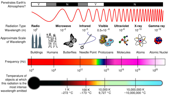
The diagram above, by courtesy of NASA, shows the basic range of electromagnetic radiation. It also shows where particular wavelengths with commonly used labels, such as "radio waves", occur on the broader spectrum. The visible spectrum, the light that the human eye perceives, is identified by the multi-coloured area near the centre of the frequency scale. At somewhat higher frequencies the radiation is known as ultraviolet. X-rays are found above the ultraviolet (UV) range.
The uppermost scale indicates whether or not the various frequencies of electromagnetic radiation penetrate the Earth's atmosphere. "Y" indicates that the relevant frequency does penetrate the atmosphere, and "N" signifies that the relevant radiation is largely blocked. Wavelengths, indicated in the second row, are expressed in terms of metres (using scientific notation). Wavelengths range from radio waves, at 103 metres, to the shorter Gamma rays, at 10 -12 metres (ie one metre divided by one thousand billion). In the third row, the approximate sizes of various wavelengths are compared to the dimensions of everyday objects. The fourth row of the diagram shows the frequencies corresponding to the various wavelengths. Frequency and wavelength are inversely proportional, so as one increases the other decreases. The bottom row shows the temperatures at which objects radiate energy at the relevant frequencies.
X-rays have a wavelength in the range of 10 to 0.01 nanometres, which corresponds to a frequency range of 30 petahertz to 30 exahertz (3 × 10¹⁶ Hz to 3 × 10¹⁹ Hz). They have shorter wavelengths than UV rays. X-radiation is sometimes known as Rontgen radiation because it was discovered by Wilhelm Rontgen. He used the term X-radiation because he realised he was experimenting with an unknown type of radiation. The machines currently used to take X-ray pictures produce X-rays with energies of around 120,000 electron volts.
X-rays are a form of ionizing radiation, and exposure to them can be a health hazard. They can penetrate solid objects, and are consequently potentially dangerous to human beings. Energy that does not pass through the body is absorbed within it and may cause permanent biological changes or damage.
A special type of photographic film, or a digital sensor, is used to record X-ray pictures. The X-rays pass through the subject and are then converted into light. Denser subjects (eg bones) absorb more energy so less energy reaches the recording medium. The relevant relevant area of an image is therefore rendered darker.